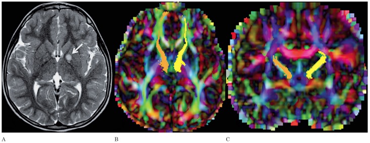
Figure 3.
A) Axial T2-weighted image of patient 3 shows a NBO in the left globus pallidus (arrow). Axial (B) and coronal (C) color-coded FA maps with superimposed FT of the left (yellow) and right (orange) anterior thalamic radiations. The NBO in the left globus pallidus does not have any effect on the adjacent anterior thalamic radiation.

