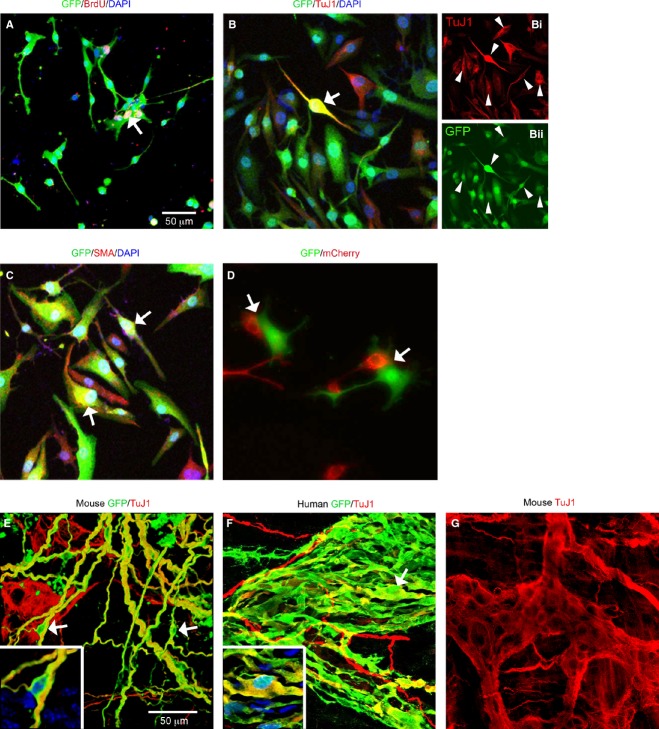
Figure 2.
Characteristics of lentivirus-transduced mouse and human gut-derived cells in vitro and following transplantation in vivo. (A) Transduced (eGFP) mouse cells proliferate as shown by BrdU incorporation (red). Arrow shows double-labeled lentivirus-transduced eGFP expressing cell also positive for BrdU. (B and C) Human cell phenotypes that were transduced by lentivirus include neural crest-derived neurons (B, arrow; Bi, arrowheads, TuJ1 in red; Bii, eGFP in green) and non-neural crest-derived cells such as smooth muscle (C, SMA, red). DAPI (blue) labels all nuclei. (D) Mixed cultures of mouse cells transduced with either eGFP (green) or mCherry (red) lentiviruses. After 2 weeks in culture, even though some cells were juxtaposed (arrows), yellow cells (expressing both expressing eGFP and mCherry) were not observed, indicating that lentivirus cross infection does not occur. (E) Transduced mouse ENS stem cell-containing neurospheres transplanted into recipient wild-type mouse gut. (F) Transduced human ENS stem cell-containing neurospheres transplanted into recipient immune deficient mouse gut. In both cases, transduced cells were observed up to 2 months (E, mouse) and up to one month (F, human) after transplantation. High eGFP expression was maintained and transduced cells integrated into the endogenous ENS network (TuJ1 immunolabeling, red). Transduced cells expressing TuJ1 are indicated by arrows and shown at high magnification in insets (also showing DAPI in blue) in both panels. (G) Untransplanted TuJ1 immunolabeled mouse gut for comparison showing enteric ganglion. Scale bars = 50 μm; A applies to A–D and E applies to E–G.

