Figure 5.
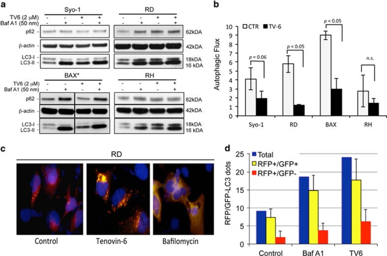
Tenovin-6 impairs autophagic flux in soft tissue sarcoma cells. (a) Synovial sarcoma cell lines (Syo-1 and BAX) and two rhabdomyosarcomas (RD and RMS) were exposed to tenovin-6 (TV6) at 2 μM concentration (*4 μM for BAX), in the presence or absence of 50 nM bafilomycin A1 (BafA1). The expression of p62 and conversion of LC3-I to its lipidated form LC3-II was determined by western blot. (b) The authophagic flux was determined by quantifying LC3-II in relation to β-actin. Autophagic flux is expressed as the ratio between LC3-II signal in the presence versus absence of Bafilomycin A1. All experiments were performed three times. (c) Autophagy flux determined by immunofluorescence using the tandem probe RFP-GFP-LC3. RD cells were transfected with the tandem probe, and Tv6 (2 μM) was added to the cultures 24 h after transfection and incubated for additional 24 h. RD cells treated with 50 nM BafA1 for 4 h were used as control for autolysosome inhibition. Autophagosome formation is visualized as yellow speckles (RFP+/GFP+), whereas autolysosomes are visualized as red speckles (RFP+/GFP−). (d) Quantification of autophagosomes (yellow) and autolysosomes (red) from 10 individual cells derived from c. The experiment was repeated twice

