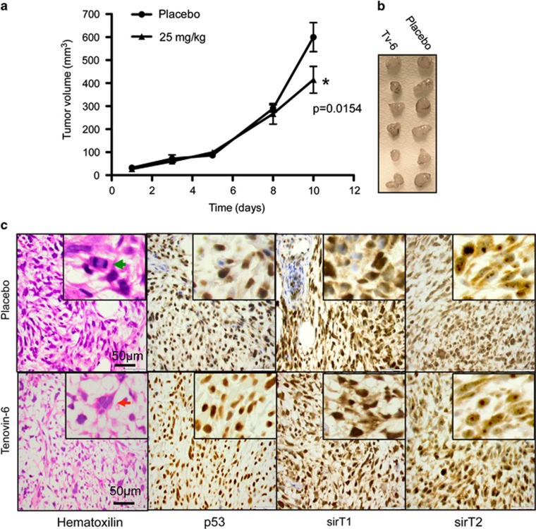
Figure 8.
Effect of TV6 on the growth of ERMS xenografts. The embryonal rhabdomyosarcoma cell line RD was xenografted in SCID mice until tumors were palpable. Mice, eight per group, were treated with Tv6 at a dose of 25 mg/kg/day, while control group were treated with vehicle. (a) Tumor volume was evaluated as described in Materials and Methods. The curve shows statistically significant differences between Tv6-treated and vehicle-treated groups (P=0.0154, Student's t-test, two sided), on the last day of treatment. (b) Excised tumors. (c) Hematoxilin/eosin staining and p53, sirT1 and sirT2 immunohistochemistry of a representative sample from control and Tv6- treated RD xenografts. SIRT1 shows cytoplasmic and nuclear staining with stronger staining in the nuclear compartment. SIRT2 staining is predominantly cytoplasmic staining with clear nucleolar accumulation of the protein. Immunohistochemical staining for p53 shows homogeneous strong nuclear staining in tumor cells from control and treated mice. Green arrow: mitotic cell. Red arrow: polynuclear giant cell

