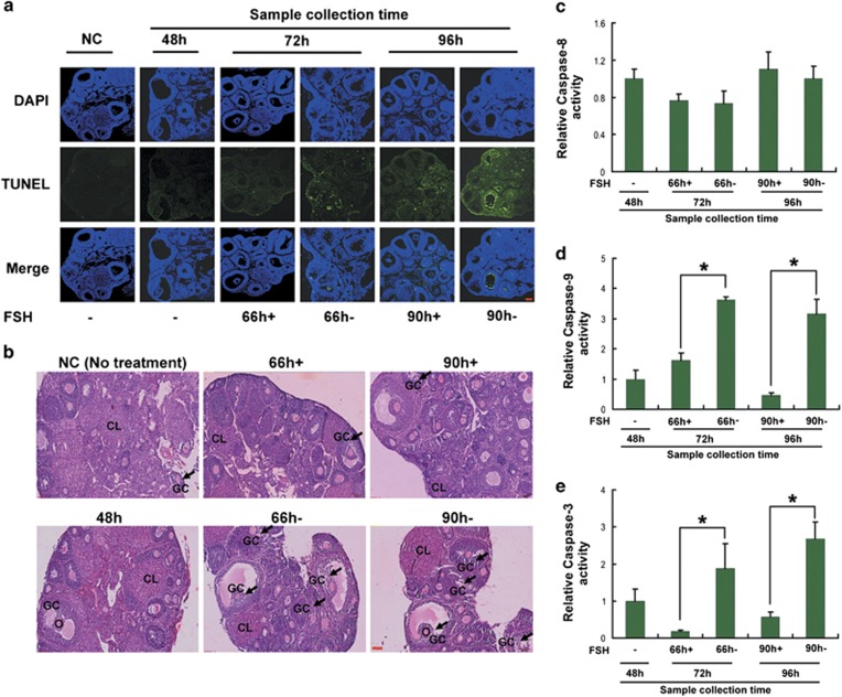
Figure 1.
FSH reduced apoptosis in MGCs of ovarian DFs. Mice were injected i.p. with FSH twice daily (12-h intervals, 08 00 and 2000 hours) for 2 days at a dose of 10 IU on day 1 and 5 IU on day 2. FSH was then withdrawn for an additional 24 or 48 h, or injected i.p. (10 IU per mouse) 6 h before MGC collection. (a–e) At 48, 72 and 96 h after the first FSH injection, the right ovaries were paraffin-embedded and serially sectioned for the TUNEL assay (a) and H&E staining (b). MGCs were isolated from DFs (>200 μm) in the left ovaries for caspase activity detection (c–e). The data represent the mean±S.E. (n=3). *P<0.05, Student's t-test. Bar, 100 μm. O, oocyte; GC, granulosa cells; CL, corpus luteum

