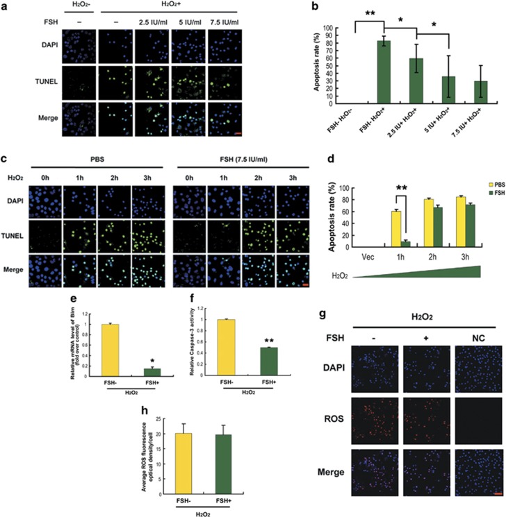
Figure 3.
FSH suppressed apoptosis in cultured MGCs. (a) Granulosa cells collected from DFs were cultured in serum-free medium containing 200 μM H2O2 and FSH at different concentrations for 6 h. The apoptotic signals were visualized with TUNEL staining (green), and the nuclei were counterstained with DAPI (blue). Bar, 20 μm. (b) Quantification of the apoptosis rates by calculating the average number of TUNEL-positive nuclei per visual field. Experiments were repeated three times, and cells were counted in three randomly selected fields from each coverslip. Data represent the means±S.E. The statistical significance between groups was analyzed with one-way ANOVA, and bars labeled with different letters differ significantly at P<0.01 (a and b) or P<0.05 (a and b). (c) After incubation with 200 μM H2O2 for 0 (vehicle), 1, 2 or 3 h, MGCs were rinsed with PBS and cultured in serum-free medium containing 7.5 IU/ml FSH for 12 h. Apoptosis was detected as above. Bar, 20 μm. (d) Quantification of apoptosis rates in each treatment as shown in (c). Data represent the means±S.E. (e–g) MGCs were incubated for 1 h in 200 μM H2O2, washed with PBS, plated in serum-free medium with 7.5 IU/ml FSH for 12 h, and then processed for qRT-PCR for Bim (e), caspase-3 activity assay (f) and intracellular ROS detection (g). Bar, 50 μm. (h) Quantification of intracellular ROS levels. The optical density was evaluated in each MGC with ImageJ 1.42q software. Data represent mean±S.E. (n=3). *P<0.05; **P<0.01, Student's t-test

