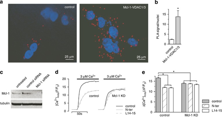
Figure 2.
Mcl-1 and VDAC1/3 interact in vivo to increase [Ca2+]mito uptake. (a) An in situ proximity ligation assay (PLA) showing the interaction between MCL-1 and VDAC1 and 3 (red) in A549 cells counterstained with DAPI (blue). (b) Quantification (mean±S.E.) of the PLA signal normalized to cell number (*P<0.01, student's t-test). (c) Western blot of A549 cells transfected with control or Mcl-1 siRNA showing efficient knockdown of Mcl-1. Depicted are representative of blots from two independent samples. (d, e) Shown in d are representative traces depicting [Ca2+]mito in A549 cells treated with control siRNA or Mcl-1 siRNAs and exposed to step increases in external [Ca2+] from 0–3 μM either in the absence or presence of VDAC-based peptides (N-ter or L14-15; 2 μM). The peak [Ca2+]mito amplitude (mean±S.E.) of the responses measured in ≥200 cells pooled from two independent samples run on two different days is shown in e (*P<0.05, ANOVA)

