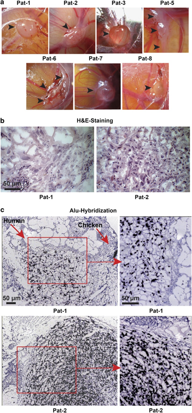
Figure 3.
GCTB stromal cells form tumor xenografts. (a) GCTB stromal cells (1 × 10⁶) derived from seven different patients were transplanted on the CAM of fertilized chicken eggs (n=8 per cell line) at embryonal development day 10 and photographed at day 17. The arrows mark the tumor xenografts. (b) H&E staining of representative frozen xenograft sections derived from Pat-1 and Pat-2 cells. (c) Alu hybridization of egg xenograft tissue derived from Pat-1 and Pat-2 cells. Dark blue-labeled cells of human origin and unlabeled chicken cells are marked by arrows. Pictures were taken at × 400 magnification, and the bar indicates 50 μm

