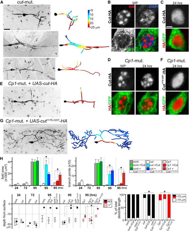
Figure 3. Cp1-Dependent Cut Isoform Production Required for Dendrite Regrowth.
(A, E, and G) Live imaging of ddaC neuron clones at 95 hr APF, with colorimetric representations of dendritic arbor depth in right panels. (A) Representative cut-mutant ddaC clones are shown. (E) Cp1-mutant ddaC neuron expressing full-length Cut is shown. (G) Cp1-mutant ddaC neuron expressing truncated Cut1176-2207 is shown.
(B–D and F) GFP and HA IHC antibody staining of ddaC neurons (ppk-Gal4; UAS-mCD8::GFP; UAS-cut-HA or UAS-cut1176-2207-HA) during metamorphosis. (B and C) Cut-HA nuclear localization patterns in control background at WP (B) and 24 hr APF (C) are shown. (D) Cut-HA nuclear patterns in Cp1-mutant clones at WP and 24 hr APF are shown. (F) Cut1176-2207-HA nuclear patterns in Cp1-mutant clones at 24 hr APF are shown.
(H) Quantitative analyses of cut-mutant dendrite regrowth defects, cut-mutant rescue experiments with full-length Cut or Cut1176-2207, and Cp1/Cut rescue experiments: field area, branchpoints, depth of primary (1°) and higher-order dendrites from surface, and percentages of total dendrite length at 95 hr APF that are shallow (within 15 μm from the body wall) or deep (≥15 μm) (n ≥ 5 in all groups). *p < 0.001, one-way ANOVA. Error bars represent SEM.
Scale bars, 25 mm (A, E, and G) and 2 mm (B–D and F). See also Figures S3 and S4.

