
Figure 2.
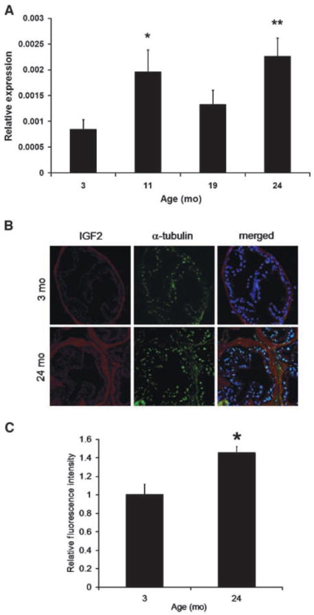
Igf2 expression increases in aging mouse DLP. A, QPCR was used to measure Igf2 expression levels in the mouse DLP of the 3-, 11-, 19-, and 24-mo-old cohorts (n = 6; **, P < 0.01; *, P < 0.05). B and C, mouse DLP sections were analyzed using immunofluorescence for Igf2 and α-tubulin (control). Images from five different random fields were acquired per section (n = 3) and the integrated density of each whole single-color image was measured with NIH ImageJ as described. Igf2 measurements were then normalized to that of α-tubulin. The older 24-mo cohort expresses significantly higher levels of Igf2 protein (P < 0.01) when compared with the 3-mo group.