
Fig. 10.
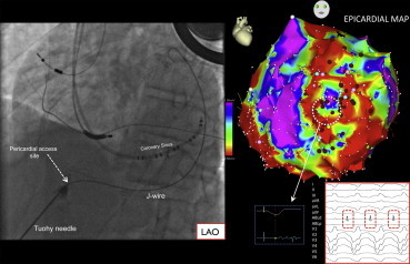
Epicardial access and mapping for ventricular tachycardia (VT). The left panel depicts an image of epicardial access in the left anterior oblique (LAO) projection. The Tuohy needed is seen entering the pericardium as indicated, small amounts of contrast are injected during the course of the needle to identify structures passed in its trajectory. A J-tipped wire passed through the needle into pericardial space outlines the lateral heart border in LAO. The right panel image depicts an epicardial voltage map in the LAO projection. A site with late potentials during baseline paced rhythm is shown (broken white circle and inset), as well as the diastolic potentials recorded during VT.