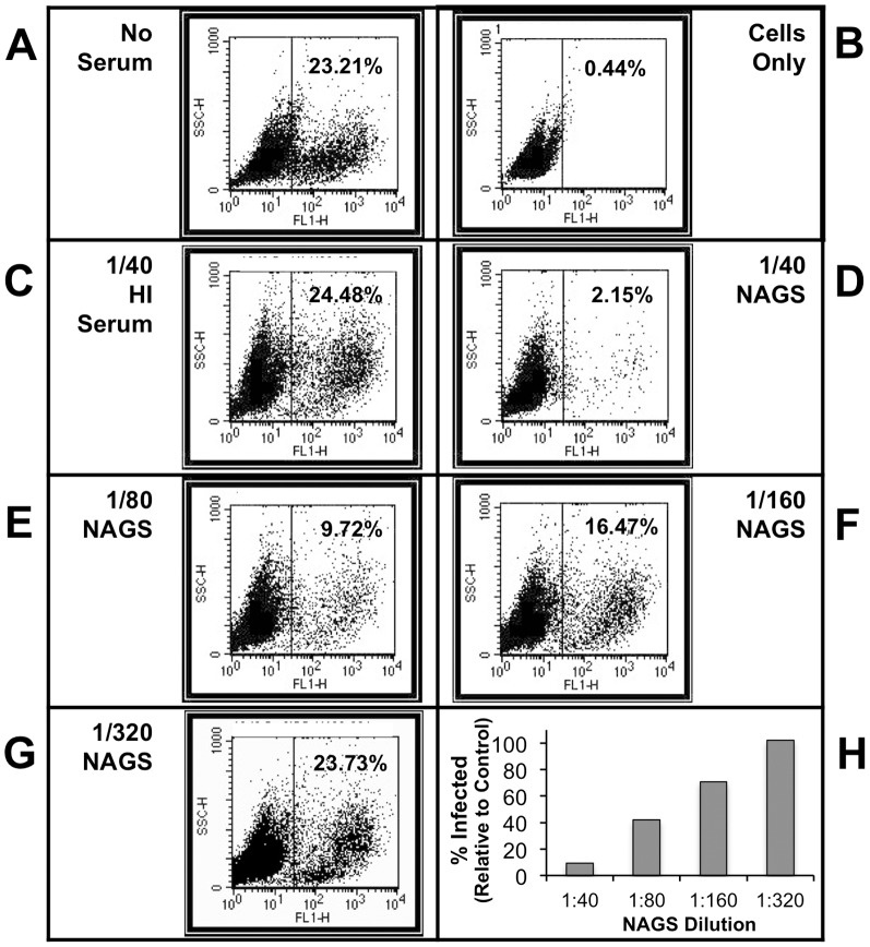
Figure 2. Flow cytometry-based PR8-GFP neutralization assay.
PR8-GFP was mixed with a range of NAGS dilutions for 40 min at 37°C, and then used to infect U937 cells for 48 hr. Flow cytometry was used to determine the percentage of the cell population that was positive for GFP expression. Controls were: (A) Virus with no serum; (B) Cells only—no serum and no virus; (C) Virus plus 1∶40 HI AGM serum. AGM serum dilutions used to treat virus were: (D) 1∶40 NAGS; (E) 1∶80 NAGS; (F) 1∶160 NAGS; (G) 1∶320 NAGS. (H) Percent of cells that were GFP-positive following neutralization with indicated dilutions of NAGS from one example animal, expressed relative to that found for the no serum control.

