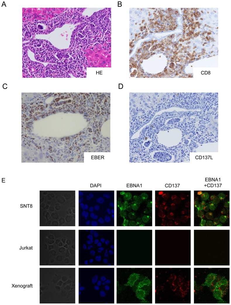
Figure 5. Histopathological specimen from the liver of the xenograft models.
We generated the models by transplanting the cells from CD8-3 patient. Nine mice were examined and the representative data were shown. (A) Hematoxylin and eosin staining showed periportal infiltration of lymphocytes. (B) Immunochemical staining with anti-CD8 antibody (brown) showed that the infiltrating lymphocytes were positive for CD8. (C) In situ hybridization of Epstein–Barr virus-encoded mRNA (EBER) (brown). Infiltration of EBV-positive cells was detected in the periportal space. (D) Immunochemical staining with anti-CD137L antibody (brown) showed that CD137L-positive cells existed in the periportal space although the number of the cells was smaller than that of EBER positive cells. (original magnification, ×400). (E) Immune-fluorescent staining with anti-EBNA1 and anti-CD137 antibodies of cells isolated from the lesions. Mononuclear cells were obtained from the tissue lesions of a model mouse, stained with the antibodies. The cells were analyzed by confocal microscopy.

