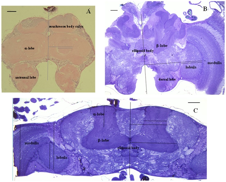
Figure 3. Brain sections and measurement of medio-lateral and antero-posterior positional data.
(A) Frontal paraffin thick section showing the α-lobe, mushroom body calyx, and antennal lobe. (B) Frontal Araldite semithin section showing the β-lobe, ellipsoid body, medulla, lobula and dorsal lobe. (C) Horizontal Araldite semithin section showing the α-lobe, β-lobe, ellipsoid body, medulla and lobula. Scale bar = 100 µm in all pictures.

