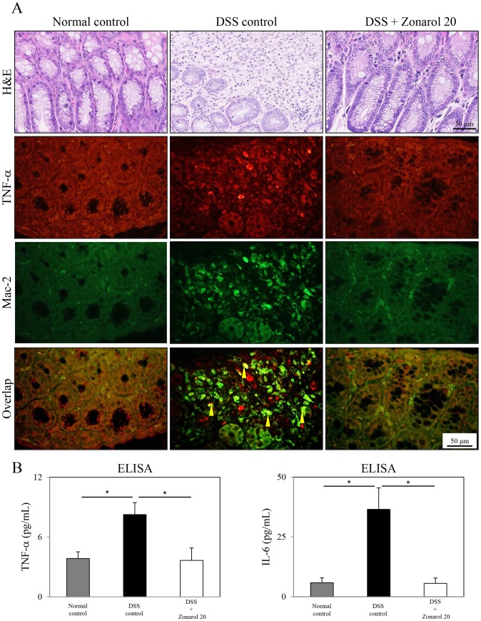
Figure 4. Zonarol suppresses the expression of pro-inflammatory signaling factors, such as TNF-α and IL-6, in mice with DSS-induced UC.
A) The IF study revealed that the number of TNF-α+ (red-stained) mucosal cells was obviously higher in the injured colons (H&E stain) of the DSS positive control mice than in the modestly inflamed colons (H&E stain) of the 20 mg/kg zonarol group (n = 6 mice per group). Additionally, the number (overlap) of both TNF-α+ (red-stained) and Mac-2+ (green-stained) macrophages in the inflamed lamina propria was also significantly more increased in the positive control mice than in the zonarol-treated mice 15 days after the DSS administration based on the IF results. Sham-treated normal control animals showed no remarkable changes, and carried no or very few cells with overlapping staining. B) Corresponding to these IHC and IF data, an ELISA demonstrated that the serum levels of not only TNF-α, but also IL-6, were significantly higher in the DSS positive control mice than in both the zonarol-treated mice and the normal control mice (n = 6 mice per group). Scale bar = 50 µm. The values are the means ± SE. *P<0.05.

