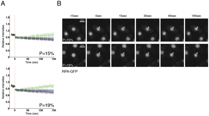
Figure 2. RPA is recruited in sites of DNA damage after laser irradiation.
(A) Cells expressing Rad11-GFP (RPA subunit) were micro-irradiated with powers 15% and 19%, and fluorescence was quantified and plotted as indicated in Figure 1A. Between 10 and 15 cells per power were analyzed. (B) Two examples of RPA-GFP micro-irradiated cells from Figure 2A are shown. Arrows indicate irradiated regions.

