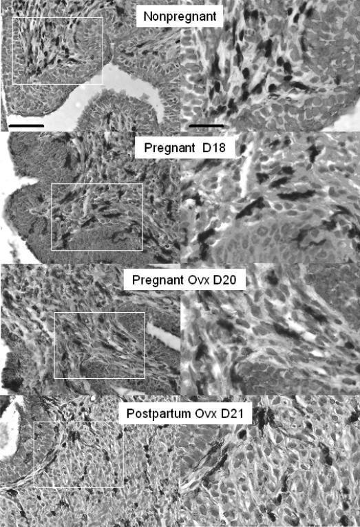
FIG. 1.
Photomicrographs of BM8 stained macrophages in cervices of Ptgfr−/− mice that were ovary intact (Nonpregnant and Pregnant D18 (day 18 of pregnancy)), ovariectomized, and pregnant (Ovx D20), or postpartum (Ovx D21). Postbreeding day is indicated. The areas within the white boxes in the left panels are magnified and presented in the right panels. Scale bar = 50 μm (left) and 25 μm (right), and applies to photomicrographs in each column. Macrophages with similar morphology and density were present in cervices from wild-type controls (data not shown).

