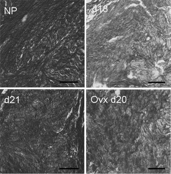
FIG. 3.
Photomicrographs of cervices from Ptgfr−/− mice at different stages of pregnancy stained with picrosirius red to visualize collagen. Coronal sections of cervices were obtained from Ptgfr−/− mice that were nonpregnant (NP), pregnant on Day 18 or 21 of postbreeding (d18 or d21), or ovariectomized on Day 20 (Ovx d20). Using polarized light, birefringence was evident as intense orange on a light yellow background before conversion to grayscale. After conversion to a grayscale image, regions of dense, highly structured collagen were dark (i.e., high birefringence with low light transmittance), whereas bright areas represent reduced collagen structure and scattered fibrils. Picrosirius red stained collagen structure to a similar extent in cervices from wild-type controls (data not shown) as to those photomicrographs shown here for Ptgfr −/− mice. Bar = 50 μm.

