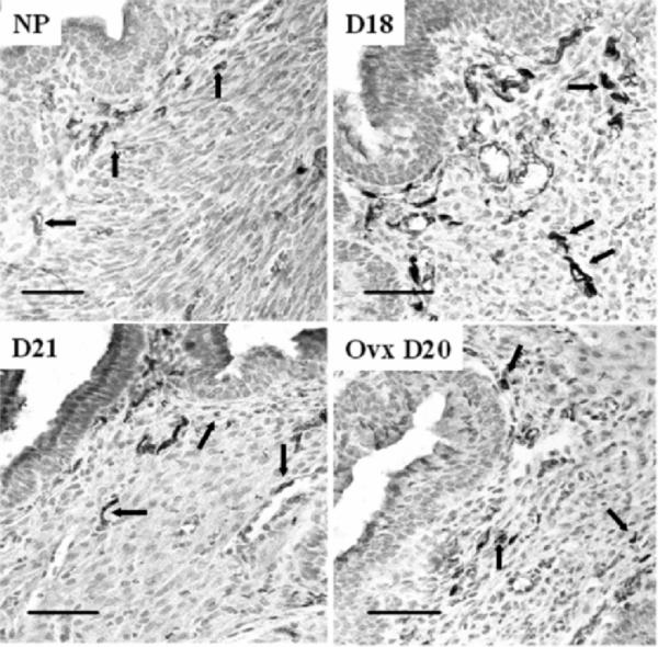
FIG. 5.
Photomicrograph of peripherin-stained nerve fibers in the cervix of nonpregnant and pregnant Ptgfr−/− mice. Groups are the same as those described in Figure 3. Luminal epithelium is in the upper left portion of each panel. Stained fibers are dark in between light gray hematoxylincounterstained cells (i.e., dark brown cells among violet-colored nuclei before conversion to grayscale). Arrows denote examples of fibers that are sparse with varicosities or cut in the plane of section in nonpregnant (NP) and pregnant D21 mice compared with the prevalent thick or bundled fibers on Day 18 of pregnancy. Morphology and density of peripherin fibers were similar in cervices from wild-type controls (data not shown). Bars = 50 μm.

