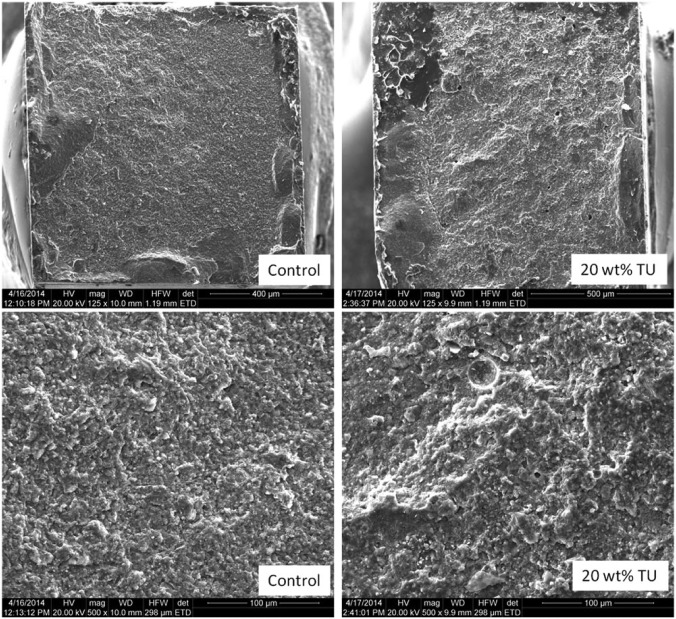
Figure 2.
Scanning electron microscope images of fractured surfaces of microtensile bond strength ceramic specimens. In general, the modified groups presented rougher surfaces compared to the control materials. However, the majority of failures were mixed adhesive-cohesive regardless of the treatment.

