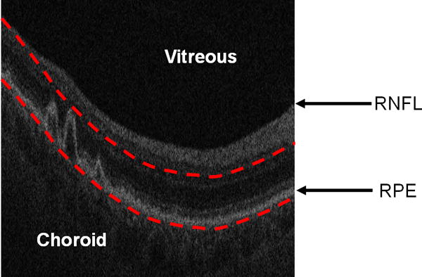
Figure 1.

Restricting the OCT image data for generating a fundus projection (the RSVP) to the vicinity of the RPE layer. The bottom red curve is the baseline of the normal RPE layer. The top red curve is determined by the largest drusen height in all of B-scans. The RSVP thus excludes extraneous portions of the retina that may add noise to the projection, such as those caused by the vitreous, retinal nerve fiber layer (RNFL), and choroid.
