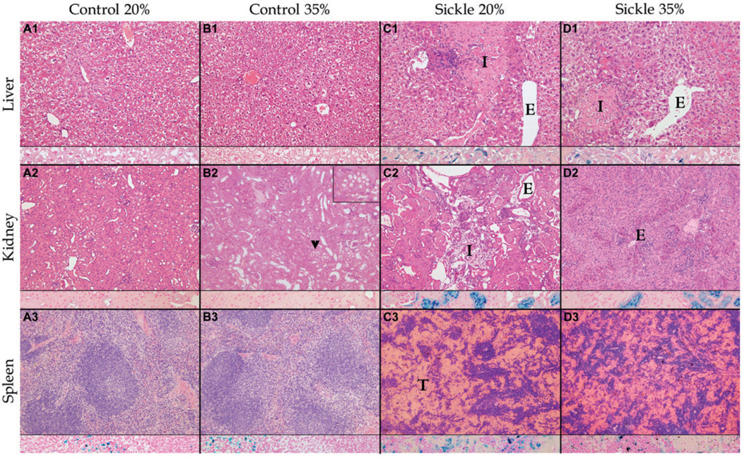
Figure 1.
Comparison of histopathological findings after 3 months of feeding in weanling sickle mice (S) receiving normal (20%) or high (35%) protein feeds with age-, sex-, and feed-matched controls (C) shows less tissue injury in sickle mice on the high protein feed (S35) compared with mice on the 20% protein feed. Columns (from left to right) show representative findings in C20 (a), C35 (b), S20 (c), and S35 (d) mice. Each column shows a section of liver (row 1), kidney (row 2), and spleen (row 3), each with an inset showing iron staining (blue) at that site. No significant differences were noted between C20 and C35, except multifocal cytoplasmic vacuoles within renal tubular epithelium (arrow heads and inset in upper right-hand corner) in C35 (b2). Both S20 and S35 are significantly different from C20 and C35 with hepatic infarcts (I) in c1 and d1; renal infarcts (I) in c2; vascular ectasia (E) in c1, c2, and d1; and thrombosis (T) of splenic sinusoids in c3 and d3. Organ injury was more severe in S20 than in S35 mice. The representative photomicrograph shows a smaller sized infarct in the liver, absence of infarct in the kidney, less distention and thrombosis of splenic sinusoids in S35 compared with S20. Micrograph was randomly selected from a collection of micrographs of each organ (Zeiss AxioSkop; hematoxylin and eosin stains [H&E], original magnification × 200, with iron [blue] stain insets, original magnification × 400); Olympus DP70 Camera; Olympus DP2 TWAIN Software

