
Fig. 4.
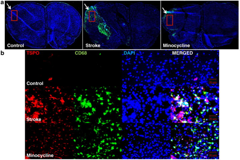
a CD68 immunohistochemistry demonstrates intense staining (arrow) indicating marked microglial activation in the regions corresponding to the infarct. Microglial activation is ameliorated by the addition of minocycline. The rectangle indicates the region from which immunofluorescence images in b were obtained. b Immunofluorescence for TSPO, CD68, DAPI, and merged images, for the controls (top), stroke (middle), and stroke plus minocycline groups (bottom) at 22 days poststroke induction. In the infarct region, large numbers of activated microglia indicated by CD68 staining coexpress TSPO, whereas they are nearly absent in the non-infarct regions and in the controls.