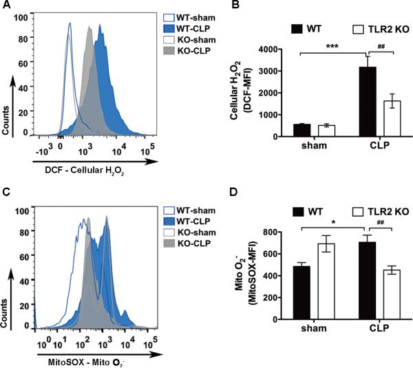
Figure 4. Absence of TLR2 attenuates leukocyte cellular H2O2 and mitochondrial O2− production during polymicrobial sepsis.
WT and TLR2−/− mice were subjected to sham or CLP procedures. Twenty-four hours later, peritoneal leukocytes were harvested, stained with either 10 μM of DCF or 2.5 μM of MitoSOX, and analyzed with flow cytometry for intracellular H2O2 (A-B) or mitochondrial O2− (C-D) production. The numbers of samples in panel B: WT-Sham, n = 4; WT-CLP, n = 5; TLR2KO- Sham, n = 5; TLR2KO-CLP, n = 5. The numbers of samples in panel D: WT-Sham, n = 17; WT- CLP, n = 35; TLR2KO-Sham, n = 15; TLR2KO-CLP, n = 23. * P < 0.05, *** P < 0.001 versus sham. # P < 0.05, ## P < 0.01 versus WT. Each error bar represents mean ± SEM. MFI = mean fluorescence intensity; DCF = dichlorodihydrofluorescein diacetate; WT = wild type; KO = knockout; CLP = cecum ligation and puncture; Mito O2− = mitochondrial superoxide; H2O2 = hydrogen peroxide.

