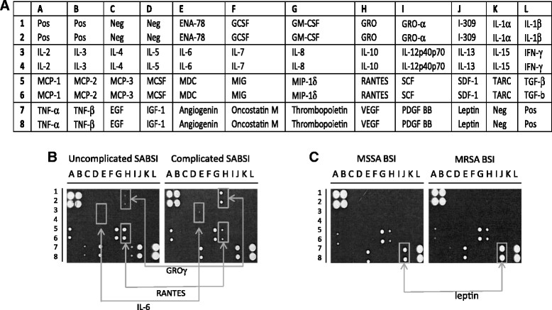
Figure 1.

Analysis of differential cytokine levels in pooled plasma from patients with SABSI. The cytokine antibody array map (RayBiotech Inc., GA) showing the positions of 42 duplicate cytokines, positive and negative controls (A). Hybridization patterns for pooled plasma from three patients per group, comparing patients with complicated SABSI to patients with uncomplicated SABSI (B) and patients with MSSA to patients with MRSA (C). Arrows indicate cytokines selected for investigation in all patients (B and C) and were based on a fold change in levels of ≥1.4 between groups. Fold change was estimated from cytokine signal intensity from compared groups (uncomplicated/complicated, MSSA/MRSA) where signal intensity was normalised with respect to the signal intensity of six positive control spots on each membrane, located at positions A1, A2, B1, B2, L7, L8.
