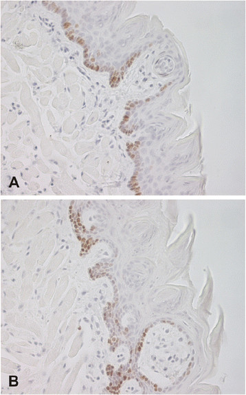
Figure 3.

Immunohistochemical staining of Ki67 proliferation marker in tongue specimens fromCar6−/−(A) and WT (B) mice. Both samples demonstrate a number of positive nuclei mainly located in the basal layer of the stratified epithelium. Original magnifications × 400.
