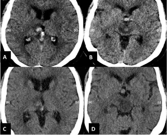
Figure 1.

Imaging before conivaptan administration. The immediate post-procedure (intra-arterial stent insertion) CT head (without contrast) demonstrate acute hemorrhage in the right mesial thalamus (A) with surrounding mild vasogenic edema and hypodensity on both sides of mid brain (B). The CT head (without contrast) before conivaptan administration reveals slight decrease in the density of acute hemorrhage in the right mesial thalamus, but with worsening surrounding vasogenic edema (C). The hypodensity in the mid brain has also worsened without any normal density tissue visible, especially adjacent to the quadrigeminal cistern (D).
