Figure 3.

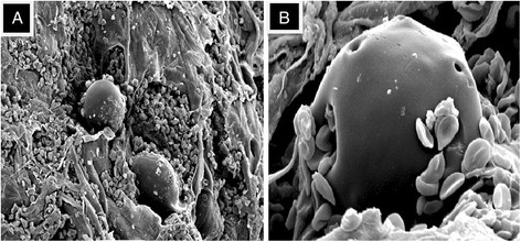
Scanning Electron Microscopy (SEM) of Arista® AH post-application. Note the presence of red blood cells and fibrin strands adhering to the Arista® AH polysaccharide hemosphere. A) Low magnification (450×). B) High magnification (1900×).

Scanning Electron Microscopy (SEM) of Arista® AH post-application. Note the presence of red blood cells and fibrin strands adhering to the Arista® AH polysaccharide hemosphere. A) Low magnification (450×). B) High magnification (1900×).