
Figure 1.
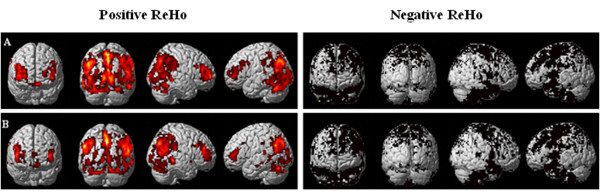
Resting-state regional homogeneity (ReHo) of one-sample t-test in baseline (A) and pain status (B) (P < 0.05, cluster threshold ≥ 10). Based on the normal topography of the selected resting-state networks, back muscle pain evoked ReHo abnormalities in the frontal lobe, parietal lobule, cingulate cortex and cerebellum. Note: Regional positive in ReHo for each network are shown as red, whereas negative are shown in black.