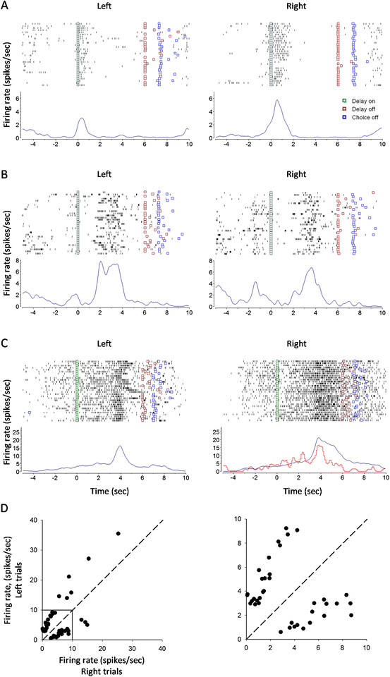
Figure 4.

mPFC neurons with differential discharge during the early, middle and late delay. (A) An early-delay neuron with firing preference for right trials (p < 0.01 for right vs. left trials; rank-sum test). (B) A middle-delay neuron with firing preference for left trials (p < 0.01 for left vs. right trials). (C) A late-delay neuron with firing preference for right trials (p < 0.01 for right vs. left trials). Red line: error trials. (D) Plotting of firing rates during left-trial delay against those during right-trial delay for the differential delay neurons (n = 47). Shown in the right panel is the enlargement of the square box in the left panel.
