
Figure 1.
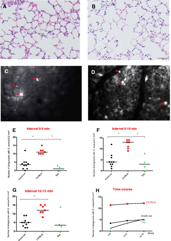
Ingestion of fluorescence-labelledS. aureusNewman and Newmaneapcells by alveolar macrophages over time in wild-type and SP-A−/−mice (n = 4–10).(A, B) Normal lung histology was observed after the experiment in the SP-A deficient animals (A) and in the wild-type animals (B). (C, D) A representative image of intravital microscopy shows bright, shining phagocytes in the alveoli which have ingested fluorescent bacteria. Clearly less of those phagocytes could be seen in SP-A deficient animals (C) compared to wild-type animals (D). (E-H) The count of such cells is presented in three subsequent intervals (E, F, G). A slight increase was observed over time (H).