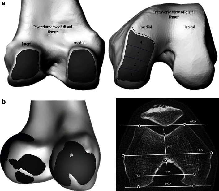
Fig. 1.
a Regions used to define and place markers on the flexion facets (left) and extension facets (right) in the distal femur to fit spheres. Boundaries of the facets were identified on CT scans; the extension facet extended until the medial sulcus terminalis (dotted line) and was split into four sections for more detailed analysis. b Visualization of the spheres fit to the flexion facets of the medial and lateral condyles, and transverse CT slice illustrating commonly defined axes for the distal femur (right)

