
Figure 3. Histology of in situ prostate cancer, 3D organoids, and xengrafts.
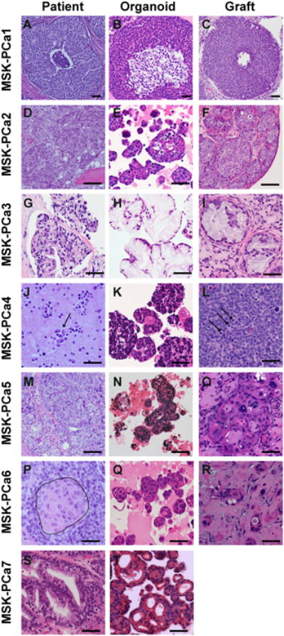
A. H&E of prostatectomy specimen of patient MSK-PCa1. B. H&E of organoids of MSK-PCa1. C. H&E of subcutaneous xenograft of MSK-PCa1. D. H&E of right acetabulum with metastatic prostate cancer from patient MSK-PCa2. E. H&E of organoids of MSK-PCa2. F. H&E of renal capsule xenograft of MSK-PCa2. G. H&E of retroperitoneal metastasis from patient MSK-PCa3. H. H&E of organoids of MSK-PCa3. I. H&E of subcutaneous xenograft of MSK-PCa3. J. Cytology of pleural effusion from patient MSK-PCa4. Arrows point to clusters of malignant cells. K. H&E of organoids of MSK-PCa4. L. H&E of subcutaneous xenograft of MSK-PCa4. Arrows point to mitotic figures. M. H&E of prostatectomy specimen of patient MSK-PCa5. N. H&E of circulating tumor cell-derived organoids of MSK-PCa5. O. H&E of subcutaneous xenograft of MSK-PCa5. P. H&E of extraprostatic prostate cancer from cystoprostatectomy specimen of patient MSK-PCa6. The field shown contains both adenocarcinoma and squamous differentiation highlighted in the dotted area. Q. H&E of organoids of MSK-PCa6. R. H&E of subcutaneous xenograft of MSK-PCa6. S. H&E of organoids of MSK-PCa7. T. H&E of organoids derived of MSK-PCa7. Scale bars represent 50 μM. See also Figure S3 for accompanying IHC.