
Figure 3.
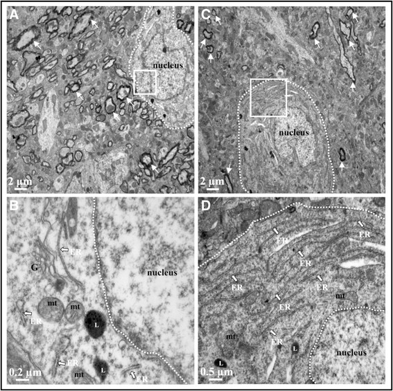
Ultrastructure of cortex after acute LPS-induced inflammation. TEM cortical images of 0.9% NaCl-injected mice (A and B) and 10 mg/kg LPS-treated mice (2 injections per 24 h, images C and D). No alteration of tissue integrity was observed in low magnification images (A and C), myelin rounded axons marked with white arrow. Images B and D represent magnified region of interest of the white square in A and C, respectively. In these magnified images, mitochondria appeared with intact cristae (mt), endoplasmic reticulum (ER) was present with ribosomes and lysosomes (L) were observed. Five sections in each area (cortex and hippocampus) were observed for each mouse brain.