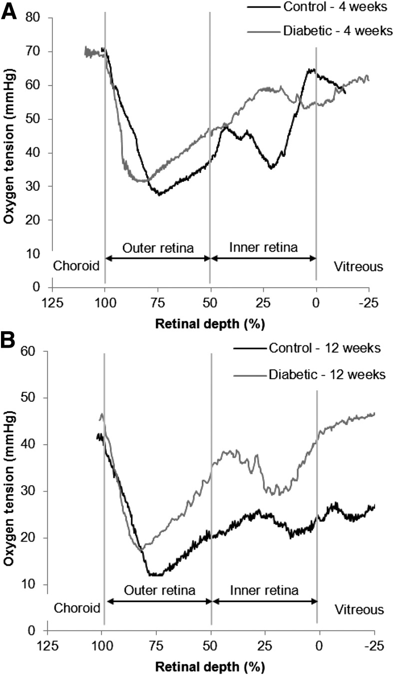
Figure 1.
Example intraretinal PO2 profiles from control and diabetic rats at 4 weeks (A) and 12 weeks (B). The choroid is located beyond 100% retinal depth. The outer retina, where the photoreceptors lie, is avascular and is located at 50–100% retinal depth. The minimum PO2 in the outer retina (Pmin) occurs in the inner segment layer. The location of Pmin in profiles can be affected by whether the electrode pulls on the retina and was not consistently different between diabetic rats and controls. The inner retina is located at 0–50% retinal depth and the vitreous is located at <0% retinal depth.

