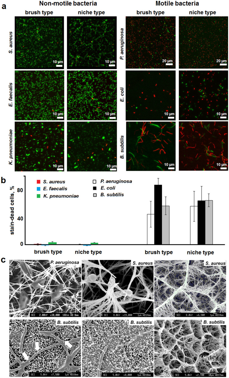
Figure 3. Bactericidal activity of nanowire titania surfaces.
(a) Fluorescence micrographs of bacterial cells incubated on different surfaces for one hour. Bacterial viability LIVE/DEAD BacLight™ assays simultaneously stain cells with intact (SYTO® 9, green) and damaged (propidium iodide, red) membranes. (b) Average number of stain-dead cells after subtracting the adhesion of S. aureus on flat titanium surfaces taken as a reference control. (c) Scanning electron micrographs of nanowire-pierced bacterial cells after one-hour incubations on both titania types (e.g. compare for B. subtilis) under dynamic conditions. White arrows point to exemplar piercing cites.

