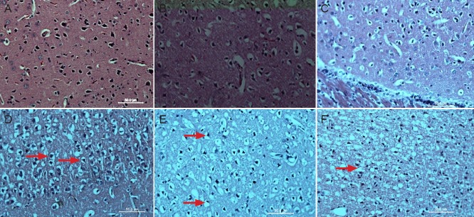
Figure 3.

Pathological changes in the cerebral infarction area in rats with cerebral ischemia-reperfusion injury (hematoxylin-eosin staining, light microscopy, × 400).
(A) Sham surgery group. Neural cells are normal looking. (B, C) 0 and 2 hours after middle cerebral artery occlusion, neural cells display no obvi-ous change. (D) 6 hours after middle cerebral artery occlusion, neural cells appeared slightly swollen (arrows). (E) 12 hours after middle cerebral artery occlusion, neural cells appeared swollen, with a bigger volume and nuclear condensation (arrows). (F) 24 hours after middle cerebral artery occlusion, no complete cell membranes are detectable (arrow).
