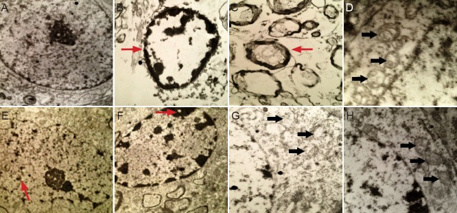
Figure 4.

Ultrastructural alterations in the cerebral infarction area in rats with cerebral ischemia-reperfusion injury (electron microscope).
(A) Normal-looking neural cells in the sham surgery group (× 8,000). (B, C) Early apoptosis (arrows) 4 hours after middle cerebral artery occlu-sion (× 8,000). (D) Mitochondrial (arrows) changes 24 hours after middle cerebral artery occlusion (× 15,000). (E) Intermediate stages of apopto-sis (arrow) 2 days after middle cerebral artery occlusion (× 8,000). (F) Pyknosis (arrow) 4 days after middle cerebral artery occlusion (× 8,000). (G) Late stage of apoptosis (arrows) 6 days after middle cerebral artery occlusion (× 15,000). (H) Partial mitochondrial (arrows) recovery 18 days after middle cerebral artery occlusion (× 15,000).
