Figure 1.

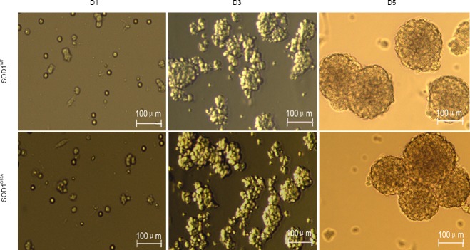
Morphology of neural stem cells prepared from embryos of SOD1WT and SOD1G93A mice at 1, 3, and 5 days after culture (D1, 3, and 5) (inverted microscope).
Embryonic neural stem cells formed typical neurospheres after in vitro culture. SOD1: Superoxide dismutase 1.
