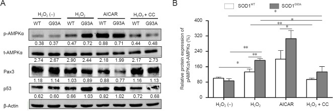
Figure 8.

Effects of adenosine monophosphate-activated protein kinase (AMPK) activator 5-aminoimidazole-4-carboxyamide ribonucleoside (AICAR) and inhibitor compound (CC) on the phosphorylation of AMPKα and protein levels of Pax3 and p53 in neural stem cells (NSCs) (western blot analysis).
(A) Representative western blots displaying the phosphorylation of AMPKα, paired box 3 (Pax3), and p53 protein in NSCs exposed to different fac-tors. β-Actin was used as a loading control. Data below each lane represent the relative gray scale compared with β-actin control bands. (B) Phos-phorylation levels of AMPKα relative to total AMPKα levels. The expression level in untreated superoxide dismutase 1 wild-type (SOD1WT) NSCs was set at 100%. Data are presented as the mean ± SD and analyzed using one-way analysis of variance followed by the least significant difference post-hoc test. p-AMPKα: Phosphorylated AMPKα; t-AMPKα: total AMPKα; H2O2 (–): Untreated; WT: SOD1WT NSCs; G93A: SOD1G93A NSCs *P < 0.05; **P < 0.01; SOD1: Superoxide dismutase 1.
