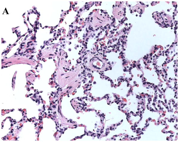
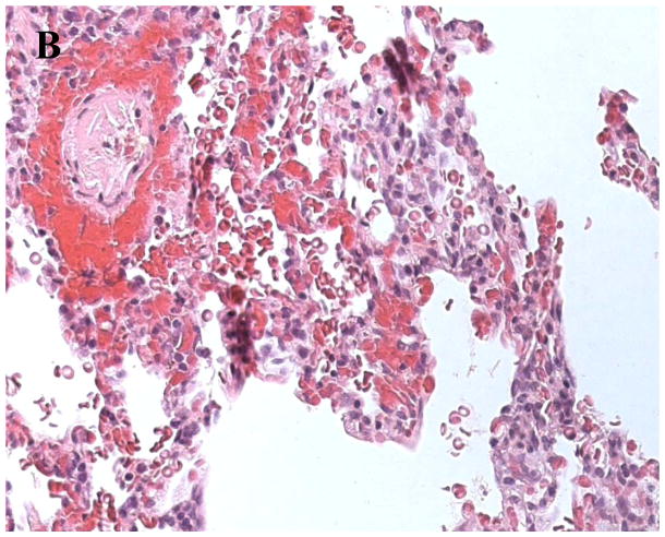
Fig. 3.
A Histological analysis of a GalTKO.hCD39 transgeneic pig lung after 4 hours of xenogeneic perfusion. Histology shows an intact pulmonary tissue with only very little hemorrhage and cell infiltration. B. Example of a hyperacutely rejected lung. The slide shows a thrombosed vessel and massive hemorrhage in lung tissue from a wild type pig after 12 minutes of xenogeneic perfusion.


