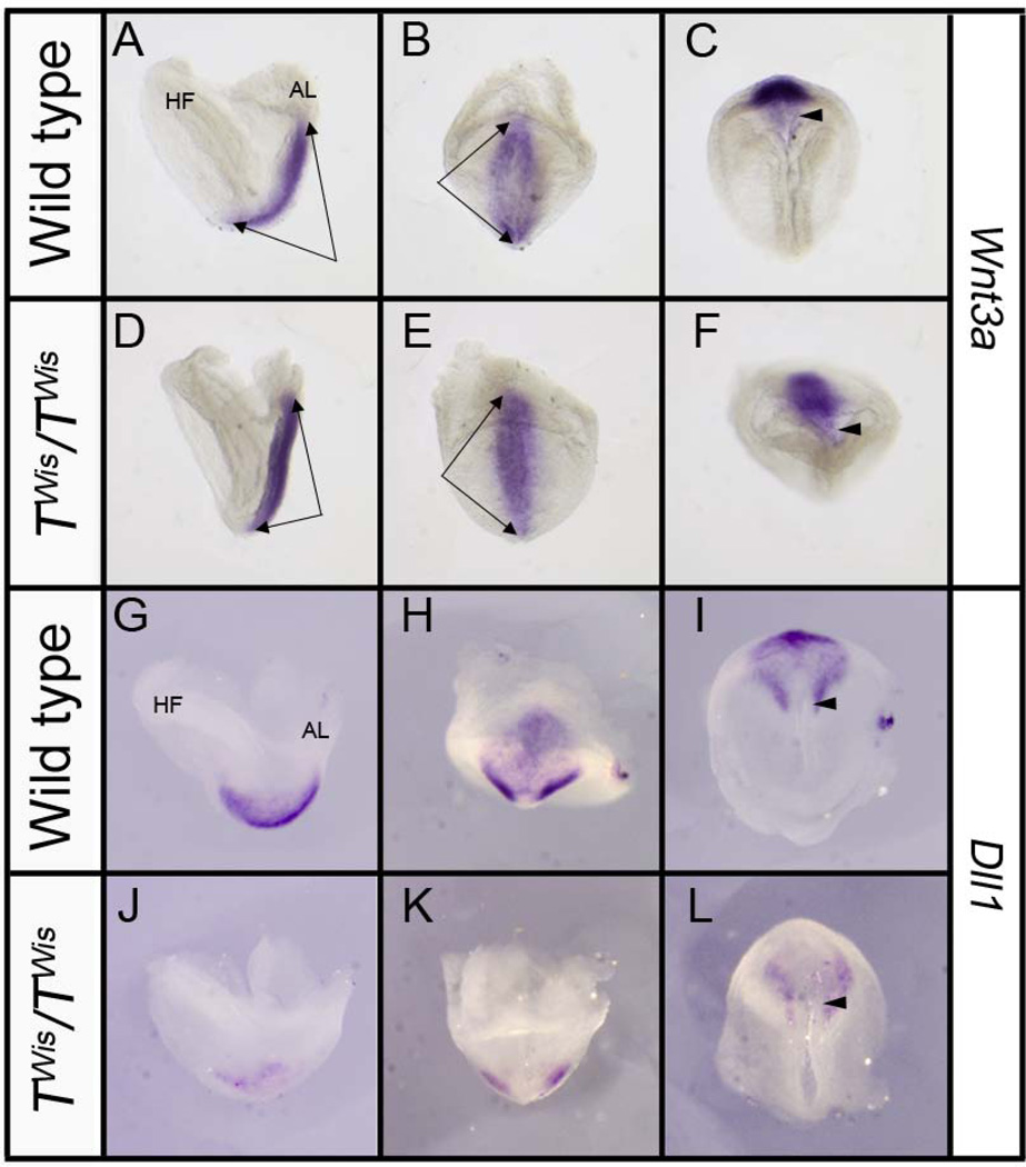
Figure 5. Expression of Wnt3a and Dll1 in wild type and TWis/TWis embryos at E8.0.
A–F: Left lateral (A, D), posterior (B, E), and ventral (C, F) views of the distal tip of wild type and TWis/TWis embryos showing similar Wnt3a expression throughout the entire primitive streak (double arrows); arrowheads point to the node in ventral views. G–L: Left lateral (G, J), posterior H, K), and ventral (I, L) views of Dll1 expression in the presomitic mesoderm and perinodal region in wild type embryos and greatly reduced expression in TWis/TWis mutants, particularly in the presomitic mesoderm. AL, allantois; HF, headfold. Arrowheads point to the node in ventral views.

