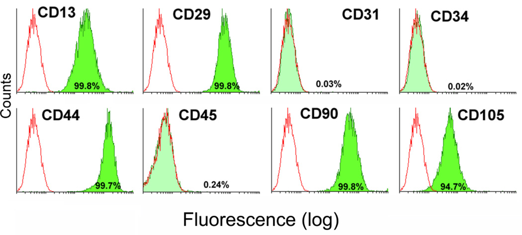
Figure 1. Analysis of stem cell surface markers on UCMSCs.
Expanded umbilical cord matrix cells (UCMSCs) that were collected from two individuals and pooled were labeled with antibodies against cell surface antigens (CD13, CD29, CD44, CD90 CD31, CD34, CD45, and CD105) and analyzed via flow cytometry. Open histograms represent the background signal; the green histogram indicates a positive signal for the indicated antibody. The percentage listed depicts the percentage of cells that showed a positive signal above background for the indicated antibody.

