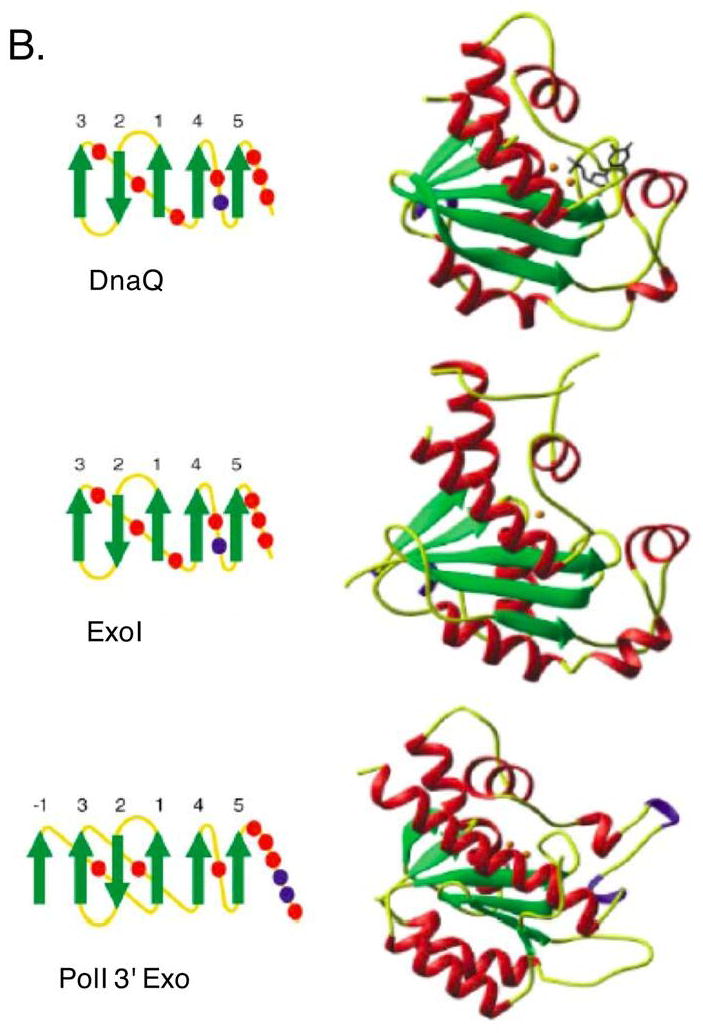
Figure 1.


A. E. coli’s DnaQ superfamily members. Shown are aligned amino acid sequences of exonuclease I, exonuclease X, oligoribonuclease, RNase D, RNase T, and the 3′ exonucleases of DNA polymerases I, II and III. Conserved acid residues are shown in bold and comprise metal coordination residues for those proteins with determined three-dimensional structure. Numbers refer to amino acid residues not shown. B. Structure of three members of the DnaQ/DEDD superfamily: DnaQ, ExoI and Polymerase I 3′ exonuclease domain. Figure republished with permission from Hamdan et al. 2002.
