
Fig. 4.
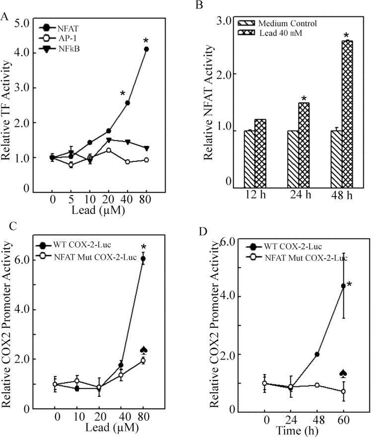
NFAT transactivation was crucial for cox-2 transcription following lead exposure.
(A), C6 cells were stably transfected with the AP-1, NFκB and NFAT luciferase reporter in combination with the pRL-TK vector (Promega) as an internal control. After the cells were treated with lead at the indicated doses, the luciferase activities were determined using a luminometer to determine transactivation activities of the individual transcription factor. The asterisk (*) indicates a significant increase compared with medium control (p < 0.05). (B), The C6 stable transfectants of NFAT luciferase reporter in combination with the pRL-TK vector were treated with 40 μM lead for 12 h, 24 h and 48 h. The transactivation of NFAT was determined using a luminometer. The asterisk (*) indicates a significant increase compared with medium control (p < 0.05). (C and D), The cox-2-luciferase reporter plasmid containing site-directed mutant of the NFAT binding sequence was used to validate the involvement of NFAT in induction of COX-2 by lead. The asterisk (*) indicates a significant increase compared with medium control (p < 0.05). The heart symbol (♠) indicates a significant decrease compared with that of wild type cox-2 promoter luciferase reporter (p<0.05).