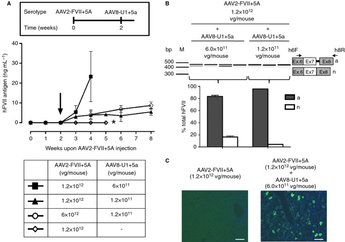
Figure 3.
U1+5a-mediated rescue of hFVII expression by AAV delivery. (A) Plasma hFVII antigen levels (mean and 95% confidence intervals) in mice (four mice per group) injected with the AAV2-FVII+5A and, 2 weeks later (arrow), with the AAV8-U1+5a viral vectors at the doses reported in the table. *Denotes the last time-point for this group of mice (n = 7). Inset: scheme of the sequential injection protocol. (B) Analysis of hFVII mRNA forms in mouse liver. The aberrant (a) and normal (n) transcripts are depicted on the right. Arrows indicate primers used for RT-PCR. Electrophoretic separation of amplicons and analysis was as in Fig. 1. Mice were injected with 1.2 × 10¹² vector genomes (vg) per mouse of AAV2-FVII+5A and 6 × 10¹¹ or 1.2 × 10¹¹ vg per mouse of AAV8-U1+5a. M, size marker; bp, base pairs. (C) Representative examples of liver sections from mice treated as indicated. Cells were stained with a species-specific anti-hFVII antibody (green) and cell nuclei (blue) with DAPI. Stained sections were captured as indicated in Fig. 1. Scale bar, 50 μm.

