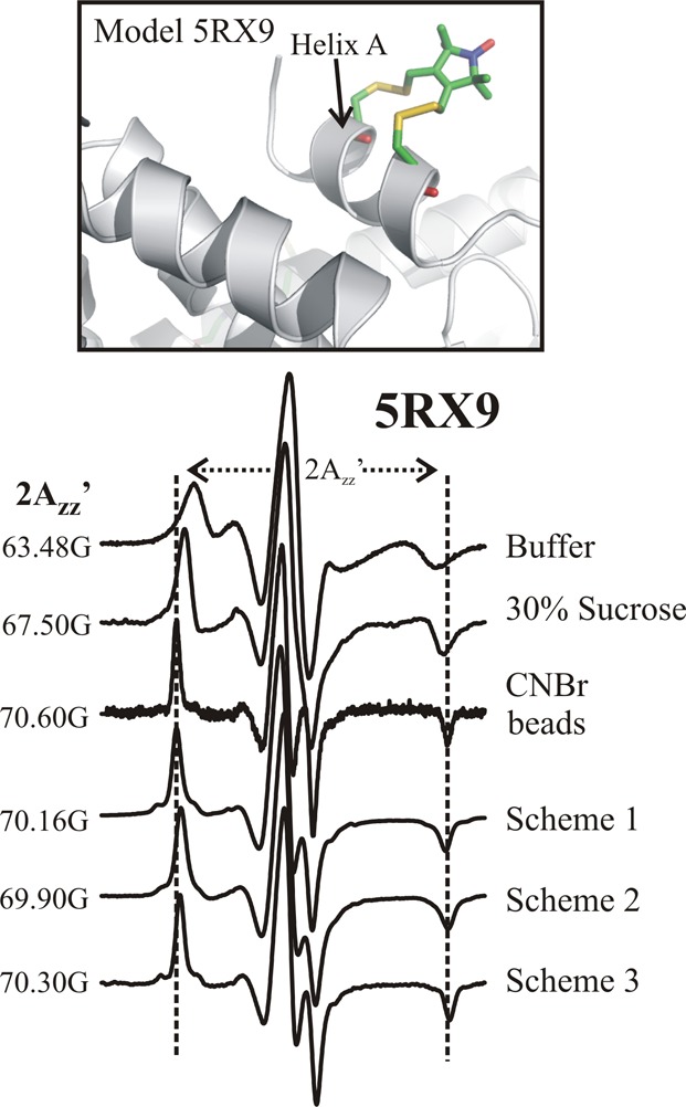
Figure 3.

Effect of tethering on protein rotational motion. (Top) Ribbon model of 5RX9 based on a crystal structure.7 (Bottom) EPR spectra of 5RX9 recorded under the indicated conditions. The vertical dashed lines define the effective hyperfine splitting (2Azz′) of T4L 5RX9 tethered on CNBr-Sepharose; the hyperfine splitting values for the spectra are given. The site-specifically tethered proteins were attached via residue 131p-AzF. The spectra for T4L 5RX9 in buffer, 30% sucrose, and nonselectively attached to CNBr-activated Sepharose via native lysine residues have been previously published and are reproduced here for reference.7
