
Figure 2.
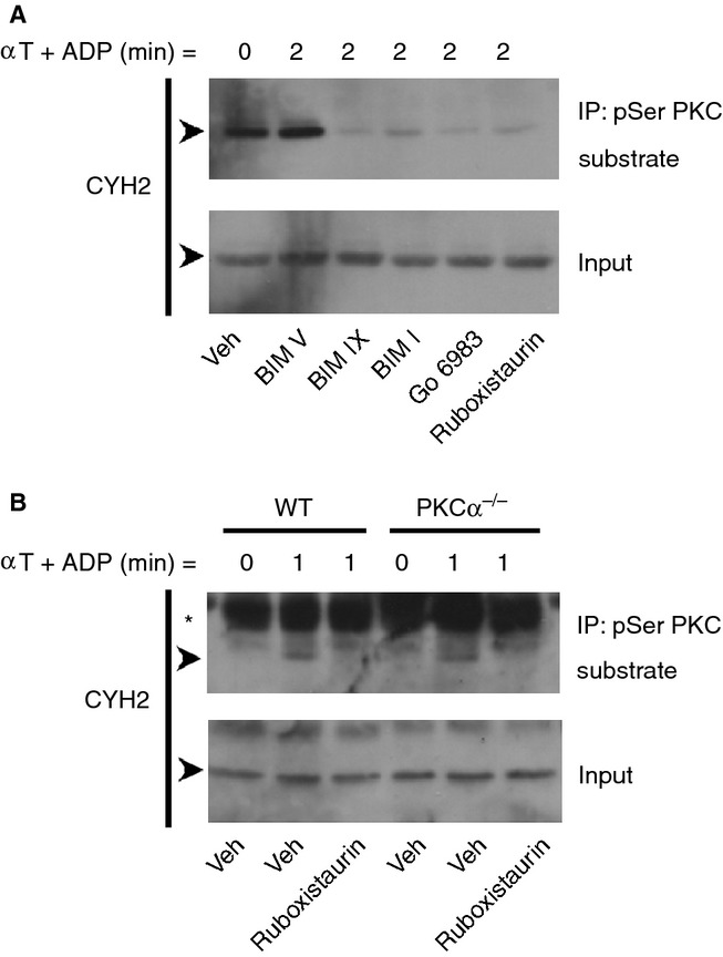
Conventional PKC isoforms mediate the phosphorylation of cytohesin-2. Washed human platelets (4 × 10⁸ mL−1) were treated for 15 min with 0.2% DMSO vehicle (Veh), the control BIM compound BIM V (10 μm), the broad-spectrum inhibitors PKC inhibitor BIM IX (2 μm), BIM I (5 μm) and Go 6983 (10 μm), or the PKCα/β selective inhibitor ruboxistaurin (10 μm) (A). Alternatively, mouse wild-type (WT) and PKCα knockout (PKCα−/−) mouse platelets (2 × 10⁸ mL−1) were used (B). Platelets were stimulated with 0.2 U mL−1 α-thrombin (αT) in the presence of 10 μm ADP and lysed at the indicated time-points. Clarified whole cell lysates and immunoprecipitations using the pSer PKC substrate antibody were immunoblotted for cytohesin-2 (CYH2). The arrows (➤) indicate the 47 kDa CYH2 band and the asterisk (*) indicates a non-specific band. Results are representative of at least three independent experiments.