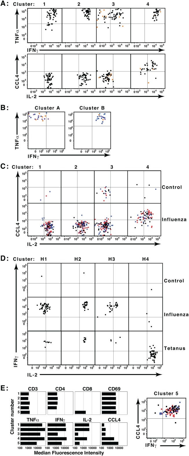
Figure 5.

Co-clustering and template assignment to rigorously compare samples. Aliquots of human PBMC were incubated in triplicate in the presence or absence of influenza or tetanus toxin peptides, then analyzed by ICS and flow cytometry. (A) Files from one unstimulated and one influenza-stimulated sample were concatenated and analyzed in SWIFT. Activated memory CD4 T cell clusters were identified by cluster gating, and the expression of four cytokines by cells in each cluster is shown. Black and red dots represent cells from the stimulated and unstimulated samples, respectively. (B) Eighteen files from triplicate samples incubated with no antigen, tetanus peptides, or four pools of influenza peptides were merged electronically into a single concatenate. Low doses of antigens were deliberately chosen so that the cytokine-producing populations in the merged sample would be very rare. The combined sample of 27.2 million cells was then analyzed in SWIFT, resulting in 1,814 split and 1,524 merged clusters. Cytokine-producing memory CD4 T cell split clusters were identified by cluster gating, and the cells in two small clusters (24 and 19 cells) are shown. Both clusters included cells from tetanus (orange) and influenza (blue) but not unstimulated samples. (C) Triplicate control and influenza-stimulated samples were assigned to the template derived in A. Triplicates are indicated by black, red, and blue dots. (D) All cells in control, influenza, and tetanus samples were assigned to a SWIFT template from concatenated tetanus- and influenza-stimulated samples. Four activated CD4 T cell merged clusters were identified by cluster gating (H1, H2, H3, and H4). (E) Clusters in the two-file concatenate (from panel A) were selected according to low staining by the live/dead marker (<1,000). Median fluorescence values are shown for all five clusters in which the ratio of influenza-stimulated to control samples was >6. These included the four CD4 T cell clusters identified in A, and one CD8 T cell cluster. [Color figure can be viewed in the online issue, which is available at wileyonlinelibrary.com.]
