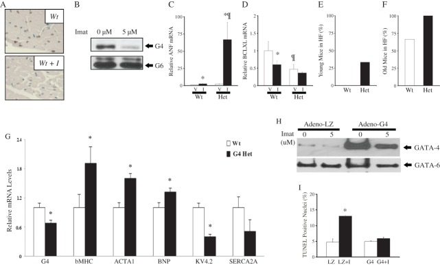
Figure 4.

A role for GATA4 in imatinib-induced cardiotoxicity. (A) Histological sections stained with a GATA4 antibody and counterstained with methyl green reveal decreased GATA4 signal (brown nuclei) in ventricles from imatinib-treated mice. (B) Western blot analysis of GATA4 protein levels in primary neonatal cardiac myocytes treated with 5 mM imatinib for 18 h. Note the decrease in GATA4 but not in the related GATA6 protein. (C and D) Graph showing ANF and Bcl-XL mRNA levels in the ventricles of wild-type (Wt) or GATA4 heterozygous mice (Het) treated with vehicle (V) or imatinib (I) as obtained by real-time PCR (QPCR). (E and F) Increased incidence of heart failure (HF) post-imatinib treatment in Gata4+/− mice (Het) as compared with their wild-type (Wt) littermates (HF = EF <45%). (G) Graphs showing transcript changes using QPCR analysis on RNA from wild type (Wt) and Gata4+/− (Het) mice ventricles. (H) Primary neonatal cardiomyocytes were infected with adenoviruses expressing GATA4 or, as a control, LacZ (LZ). Western blot showing GATA4 and GATA6 levels in the indicated conditions. (I) Quantification of TUNEL (terminal deoxynucleotidyltransferase-mediated dUTP end labelling) assay. The data are the mean of triplicate plates with 10 fields counted per plate.
